Disclaimer

Disclaimer & Warning: The information in this blog is only provided for informational purposes. This information is not designed to be used to treat any disease or health problem. Instead, always consult with your physician for proper treatment.

Wednesday, October 23, 2013

Cancer and Lactic Acid Cycle

In order to produce energy, your cells carry out cell respiration within the mitochondria, which are the power generators of a cell, converting oxygen and nutrients (glucose, amino acids and fats) into chemical energy known as adenosine triphosphate (ATP).

With this process your cells produce 38 molecules of ATP; and, then, your cells use this ATP to perform its daily functions.

Cell Respiration


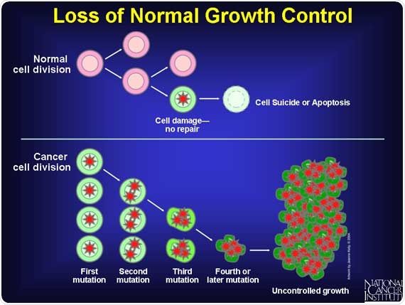
Why Cancer Patients Waste Away
At least 50% of all cancer patients suffer from a wasting syndrome called cachexia. Affected patients lose weight, including muscle, no matter how much they eat. The wasting is the immediate cause of about one third of all cancer deaths.

It appears that the cancer cells intervene and consume the majority of the glucose and other nutrients while the normal cells starve. After the cancer cells convert the glucose to energy, the cells secrete lactic acid, which is sent to the liver which converts it back to glucose and returned to the cancer cells, which continue to divide and grow out of control.

In addition, the cancer cells secrete a protein (called IMPL2), which  prevents healthy normal cells from responding to insulin, the hormone that stimulates cells to import sugar and burn it for energy. When levels of IMPL2 rise, fat, muscle and other tissues can no longer consume sugar and begin to waste away. Lowering IMPL2 levels reduces the amount of wasting.

Those stark numbers have spurred research into what exactly causes cachexia in patients with cancer and how it might be avoided with a new drug.

The good news is that there are natural substances that can block the production of lactic acid and break the cycle. See below for details.


Cachexia Cycle (Lactic Acid Cycle)
As previously mentioned, the cancer cells (and the microbes inside the cell) interrupt the glucose coming into the cell and consume (ferment) most of the glucose without oxygen. As part of this (inefficient) fermentation process, the cancer cells produce less energy along with lactic acid. 

Fermentation allows the cancer cells to produce energy without the need for oxygen. But, the cancer cells are very inefficient at processing glucose -- only about 5% compared to a normal cell. Instead of producing 38 molecules of ATP, each cancer cell (which is now acidic) produces only 2 molecules of ATP, meaning that the cancer cells are wasting a lot of energy. 

This wasted energy causes the cancer patient to become tired and malnourished. This excessive use of glucose by a cancer cell is actually part of the process whereby cancer cells actually “steal” glucose from normal cells (cancer cells also steal nutrients from normal cells). This means that normal cells can literally starve to death, creating malnutrition, pain, other health complications and eventually death.

In addition, as a byproduct of the fermentation process, the cancer cells dump lactic acid back into the bloodstream.

Then, the lactic acid is sent to the liver where the liver converts the lactic acid into glucose.

Then, the liver releases the glucose into the bloodstream where cancer cells are likely to pick up this glucose because cancer cells consume about 15 times more glucose than normal cells.

While this may seem like a harmless cycle, there are three reasons why almost half of all cancer patients die from this cycle.

First, the conversion of glucose to lactic acid by the cancer cells and the conversion of lactic acid to glucose in the liver both consume massive amounts of energy.

Second, the lactic acid itself, while it is in the bloodstream, can block key nutrients from reaching healthy (non-cancerous) cells.

Third, the cancer cells secrete a protein (IMPL2), which  prevents healthy normal cells from responding to insulin and being able to import sugar and burn it for energy.

To summarize, cachexia is the wasting away of the cancer patient’s body. The cancer metabolizes glucose inefficiently, turning it into lactic acid, which is converted back to glucose by the liver. This process consumes an enormous amount of the body’s energy, causes pain and tires out the cancer patient. This happens over and over again as the cancer grows and the rest of the body wastes away.

Summary
To summarize this process and the cycle:
  1. The cancer cells ferment massive amounts of glucose, which consumes energy,
  2. They process the glucose with fermination, which is very inefficient,
  3. A byproduct of this fermentation is lactic acid,
  4. This lactic acid then goes into the liver,
  5. The liver then converts this lactic acid back into glucose, consuming even more energy.
  6. The cancer cells secrete a protein that prevents normal cells from being able to absorb glucose.
  7. Much of this glucose is consumed by the cancer cells and the cycle starts over.
Cachexia Pathology
Cachexia is seen frequently with cancer, but is also seen with diseases such as AIDS/HIV, heart failure, emphysema, and kidney failure. With regard to cancer, it is seen most frequently with lung cancer, pancreatic cancer, and stomach cancer.

According to the National Cancer Institute, cachexia is estimated to be the immediate cause of death in 20% to 40% of cancer patients, with failure of the respiratory muscles as a frequent cause of death.  In addition, about eight out of every ten patients with advanced cancer will suffer from this potentially deadly wasting syndrome.

Cachexia not only worsens survival for people with cancer, but it interferes with quality of life. People with cachexia are less able to tolerate treatments, such as chemotherapy, and often have more side effects. For those who have surgery, postoperative complications are more common. Cachexia also worsens cancer fatigue, one of the most annoying symptoms of cancer.

Cachexia peels away both the visible muscle (somatic muscle) and the invisible muscle (the visceral proteins in the gut and elsewhere).  And because the visceral muscle serves as a reservoir for certain immune-enhancing nutrients, the loss of this muscle leads to a weakening of your immune system, making you more prone to life-threatening infections such as pneumonia.

To further complicate matters, many patients suffer from anorexia (a moderate to severe aversion to food) at the same time that they have cachexia. As a result, cancer patients may lose up to 80% of body fat and skeletal muscle. Muscle loss leads to weakness, immobility of patients, more infections and poorer response to treatment. Emotionally, cachexia promotes feelings of fatigue, depression and worthlessness. 

A number of factors often converge to cause catabolic wasting. Malnutrition due to reduced food consumption or impaired nutrient absorption occurs frequently in later stages of chronic disease and can cause marked loss of muscle and fat tissue. Even though cachexia is typically accompanied by loss of appetite, it rarely responds to increased food intake alone (Siddiqui 2006; Solheim 2013). Dehydration is another important contributor, as loss of fluid results in reduced weight (Morley 2006).

Inflammation also plays a major role in deterioration of body mass among individuals with cachexia (Morley 2006). Both acute and chronic illness can cause marked increases in the production of inflammatory cell-signaling molecules called cytokines. These inflammatory mediators alter numerous metabolic processes, resulting in reduced muscle protein synthesis and increased muscle protein breakdown.

Several specific cytokines have been linked to cachexia including interleukin-1, interleukin-2, interleukin-6, interferon-γ, and tumor necrosis factor-alpha (TNF-α). Inflammatory cytokines activate a major metabolic regulator called nuclear factor kappa B (NF-κB), which in turn drives several physiological changes that promote tissue deterioration.

Inflammatory cytokines also stimulate the release of the adrenal hormone cortisol and neurotransmitter hormones called catecholamines; both cortisol and catecholamines can exacerbate catabolic wasting by disrupting muscle cell metabolism and altering the basal metabolic rate (Siddiqui 2006; Morley 2006).

Reductions in levels of testosterone and insulin-like growth factor-1 (IGF-1) are thought to play an important role in catabolic wasting as well. Both testosterone and IGF-1 exert anabolic actions in muscle tissue, so declining levels of these hormones can lead to reduced muscle mass (Morley 2006).

Note 1: Be careful with glucose-rich intravenous feeding. This feeds the cancer more than it feeds you. Moreover, overfeeding of glucose/dextrose can lead to liver and respiratory problems. A Harvard doctor, George Blackburn, who has studied nutrition in cancer for several decades, recommends a tailored prescription of macronutrients and micronutrients of 30% lipids, including a minimum of no less than 4-6% Omega-6 fatty acids and some Omega-3 fatty acids, now known to counter wasting in cancer. Also, please keep in mind that intravenous feeding, being invasive, offers a route for blood-borne infections. 
Note 2: Before now, cachexia, characterized by muscle wasting and dramatic weight loss, was believed to spare the heart. But an Ohio State University study showed that the condition reduces heart function and changes the heart muscle structure in mice with colon cancer. The study results support the idea that insufficient heart performance might also be responsible for fatigue symptoms, leading to less exercise and more severe muscle wasting. The study is published in an issue of the International Journal of Oncology.

How to Stop Cachexia
Recommendations from many cancer organizations encourage patients to eat whatever they want of the typical American diet; that is, more saturated fats, refined flours and sugars.  But, all that does is feed the inflammation and fuel this muscle-wasting process and make the cachexia even worse!

Stopping or fixing cachexia is not a matter of simply eating more calories, from fats, carbs or protein.  Rather, the disorder is a metabolic dysfunction driven by a chronic, low-grade pro-inflammatory condition with the unrelenting and consequent breakdown of muscle and other lean tissues. 

Various biofactors have been identified as mediators of tissue wasting in cachexia. As previously mentioned, these include cytokines such as tumor necrosis factor-α (TNF-α), interleukin-6 (IL-6), interferon-γ (IFN-γ) and leukemia inhibitory factor (LIF).

Additional factors include tumor-derived factors such as lipid mobilizing factor (LMF) and protein mobilizing factor (PMF), which can directly mobilize fatty acids and amino acids from adipose tissue and skeletal muscle respectively.

It appears that anti-inflammatory supplementation like Omega-3 EFAs (especially eicosapentaenoic acid (EPA)), turmeric/curcumin, ginger,  and others, can make a pronounced impact on stopping and reversing this distressful disorder.  And, in spite of the fatigue that many patients will experience, gentle resistance exercise is essential to maintain and rebuild fragile muscles.

A sound plant-based nutritional strategy will not only help curtail inflammation, but reduce free-radical damage, minimize platelet activation (which can lead to dangerous blood clotting), manage blood sugar surges, and reduce serum levels of insulin-like growth factor 1 (IGF-1), which stimulates cell multiplication and inhibits cell death.

In addition, some studies have indicated that MSM and Vitamin C were able to reduce lactic acid; and, a substance called hydrazine sulfate was able to break the cachexia cycle. This was accomplished by the hydrazine sulfate being able to block a key enzyme in the liver to prevent lactic acid from getting converted back into glucose.

Athletes and Lactic Acid
Any athlete is familiar with lactic acid. Athletes normally take pickle juice, D-Ribose, MSM (methylsulfonylmethane) and Vitamin C or other special things to get nutrients past the lactic acid blockade and thus get energy into the cells.

Most cancer patients do not die from the cancer cells, rather they die from the damage to the non-cancerous cells. Thus, it is critical to get nutrients past the lactic acid blockage to nourish the non-cancerous cells immediately!!

Every cancer patient who thinks they may have cachexia, no matter what protocol they are on, should start taking D-Ribose immediately. Ribose is an essential ingredient in the formation and conservation of ATP, ADP and AMP (energy molecules).

Note:  
ATP = Adenosine triphosphate (3 phosphates attached)
ADP = Adenosine Diphosphate (2 phosphates attached)
AMP = Adenosine monoposphate (1 phosphate attached)


Concerning the buildup of lactic acid, many athletes prior to a strenuous workout, and after the workout, will consume MSM (methylsulfonylmethane) with Vitamin C. This combination is known to neutralize the lactic acid buildup after the workout and it stops the pain.

Using the MSM and Vitamin C together, as they work synergistically, should have the same ability to neutralize the lactic acid in patients with cachexia as well. MSM and vitamin C can be purchased at the health food store. Some MSM products, such as the Trimedica brand, have Vitamin C in the capsule along with the MSM and should work well.

Hydrazine Sulfate
Hydrazine sulphate breaks this lactic acid cycle by blocking a key enzyme in the liver to prevent lactic acid from getting converted back into glucose.
http://www.alkalizeforhealth.net/cancerpain.htm

Note: Of all of the alternative treatments for cachexia, perhaps Hydrazine Sulfate is the best known. The reason is that it was designed specifically for cachexia.

Hydrazine sulfate is the salt of hydrazine and sulfuric acid. Known by the trade name Sehydrin, it is a chemical compound that has been used as an alternative medical treatment for the loss of appetite (anorexia) and weight loss (cachexia) which is often associated with cancer.

Hydrazine sulfate has not been approved in the United States as safe and effective in treating any medical condition, although it is marketed as a dietary supplement. It is also sold over the Internet by websites that promote its use as a cancer therapy. The active ingredient is hydrazine, and the sulfate component is present to aid in formulation.

Hydrazine Sulfate works on stopping the cycle just mentioned. Hydrazine Sulphate, or more commonly Hydrazine Sulfate, interrupts the ability of the liver to convert lactic acid from tumors into glucose thereby helping to starve the tumors and inhibit their ability to metastasize.

Overall gluconeogenesis is stimulated when cancer is present. Gluconeogenesis requires a great deal of energy and excessive gluconeogenesis is thought to be a significant factor that contributes to cancer cachexia (Gold, 1968). Dr. Joseph Gold recognized in the 1960’s that metabolic strategies that inhibited the enzyme phosphoenol pyruvate carboxykinase (PEP-CK) would reduce gluconeogenesis and decrease the severity of cachexia (Gold, 1968). Dr. Gold after testing a series of compounds found that hydrazine sulfate could effectively reduce excessive gluconeogenesis in cancer (Gold, 1974, 1981).

By stopping the liver from converting the lactic acid into glucose breaks the cachexia cycle.

Dr Joseph Gold looked at the chemical process of glycogenesis and determined that, if he inhibited the PEP CK enzyme (much too large a word for anyone to try to pronounce), he could stop the process. Voila, he came up with hydrazine sulfate, a substance that is made cheaply, simple to use, and shrinks tumors. In his early animal studies, Dr Gold showed that, in greater than fifty percent of cancerous animals, he was able to stop the process of glycogenesis, end the cachexia, and the animals began gaining weight. With sugars cut off to the tumor, the tumors began shrinking.
http://www.mnwelldir.org/docs/cancer1/altthrpy2.htm


Warning! Hydrazine Sulfate may raise your blood pressure and heart beat dramatically and cause the worst headache you’ve ever experienced. This is a very dangerous condition, especially for someone already battling cancer.

Hydrazine Sulfate is an MAOI (Momoamine Oxidase Inhibitor). What it does is inhibit an enzyme that breaks down monoamines (serotonin, norepinephrine, and dopamine), those brain chemicals that make us happy. MAO inhibitors have been used as antidepressants. However, MAOs have another job in the body: they metabolize tyramine, an amino acid. When taking an MAO inhibitor, tyramine is not broken down, and eating foods with tyramine can raise your blood pressure and heart beat dramatically and cause the worst headache you’ve ever experienced.

Most of the foods containing tayramine are not on most cancer diet plans, so you should be avoiding them anyway.

Foods containing tyramine are (mainly) aged, fermented, or pickled, such as most cheeses (except cottage cheese, cream cheese, and fresh Mozzarella), lunch meats, hot dogs, yogurt, wines and beers. Here is a pretty good list of foods that contain tyramine:

In general, any high protein food that has undergone aging should be avoided. Also, any over-the-counter cold or allergy remedy should also be avoided.
http://www.mnwelldir.org/docs/cancer1/altthrpy2.htm

Here is the warning from Walter Last:
Hydrazine sulphate is a monoamine oxidase inhibitor and the following should not be used during hydrazine therapy: tranquilizers or sedatives in doses greater than 100 mg per day, especially benzodiazepines and phenothiazines should be avoided, also antihistamines, alcohol and other agents that depress the central nervous system such as morphine. Also vitamin B6 should not be taken. Foods high in tyramine must be avoided. These are aged and fermented products such as most cheeses, cured meats or fish, sour cream and yoghurt, tofu and tempeh, bouillon cubes, sauerkraut, pickles and yeast extracts. Also restricted are broad beans, avocados, bananas, raisins, figs, dates and dried fruit in general as well as overripe fruit.
http://users.mrbean.net.au/~wlast/cancer6-remedies.html

Here is the warning of Dr. Gold, developer of Hydrazine Sulphate:
HS is an irreversible and potent MAO (monoamine oxidase) inhibitor, a class of compounds that can have potentially deadly interactions with other drugs. For over three decades it has been known that central nervous system depressants—such as barbiturates, tranquilizers and alcohol—are incompatible with MAO inhibitors and use of the two together could result in extremely dangerous effects.
Note: For  details about using Hydrazine Sulfate, refer to the Hydrazine Sulfate Protocol on the Cancer tutor website:

Cesium Chloride
One option to help hydrazine sulfate stop this lactic acid cycle is alkalinity. Cesium chloride (and a few other minerals), the most common substance to make cancer cells alkaline in an alkaline treatment program, has been proven by Dr. A. Keith Brewer, PhD, to get into cancer cells, when other nutrients cannot. The cesium chloride:
-- Makes the cancer cell alkaline,
-- Limits the intake of glucose into the cell (thus starving the cell),
-- Neutralizes the lactic acid (which is actually what causes the cell to multiply uncontrollably and eventually kills the cell) and makes it nontoxic, and
-- Stops the fermentation process, which is a second affect of limiting the glucose (fermentation is what creates lactic acid in the first place).

In other words, cesium chloride will break the cycle in several different ways.

But this is the important point: Hydrazine Sulfate blocks the cachexia cycle in the liver and cesium chloride blocks the cachexia cycle in the cancer cells.

Both cesium chloride and hydrazine sulfate are complex treatments and have many restrictions.

Refer to the the following website for more details:
http://www.cancertutor.com/hydrazine/

Other Website References About Cachexia

Cachexia (Lactic Acid Cycle)
http://www.canceractive.com/cancer-active-page-link.aspx?n=3429 
http://www.clinicalcorrelations.org/?p=1285
http://www.tpims.org/disease-research/wasting-syndrome
http://mobile.the-scientist.com/article/42601/insulin-interference-triggers-cancer-linked-cachexia
http://msccc.com/cancer-resources/nutrition-in-cancer-care
http://www.doctorsacrossborders.mu/diseases/item/613-cancer-and-cachexia-silent-killer-wasting-disease.html

Tuesday, October 22, 2013

Cancer and Oxidative Stress

Your body constantly reacts with oxygen as you breathe and your cells produce energy. As a consequence of this activity, highly reactive molecules are produced within our cells known as free radicals and oxidative stress occurs.

Oxidative stress can occur through overproduction of free radicals and the unregulated production of cellular oxidants damages DNA, causing mutations and modification of gene expression. These free radicals (e.g. ROS) activate signal transduction pathways, leading to the transcription of genes involved in cell growth regulatory pathways, setting the stage for cancer development. This is indicated by, for example, high levels of oxidative lesions in cancer tissue, and reduced cancer incidence in populations with high dietary antioxidant intake.

When our protein-controlled (anti)-oxidant-response doesn’t keep up, oxidative stress causes oxidative damage that has been implicated in the cause of many diseases (including cancer) and also has an impact on the body’s aging process.

Oxidative stress, along with chronic inflammation, has been demonstrated to fuel tumor development cancer cell proliferation, invasion, angiogenesis, and metastasis by activating various oncogenic transcription factors.

It appears that some tumors (adenomas and carcinomas) have increased levels of different markers of oxidative stress, such as increased levels of ROS, nitric oxide (NO), lipid peroxides,  and low glutathione levels.

Besides lipid modifications, there is also increased leukocyte activation in carcinogenic tissue, which indicates possible contribution of inflammatory cells to a further oxidative stress and DNA damage.

Consequently, oxidative stress plays a role in the etiology of most cancers, along with the patient’s lifestyle habits (smoking, drinking, use of antioxidants, exercise, etc.). Consistent with that, antioxidant enzymes have been demonstrated to suppress tumorigenesis when being elevated both in vitro and in vivo, making induction of these enzymes a more potent approach for cancer prevention.  

Cancer and Inflammation

In Latin, the word "inflammation" means "Ignite, set alight" and like gasoline, that's exactly what it does to cancer. A microenvironment of chronic inflammation can increase the risk of cancer, bolster chemotherapy resistance and turn on oncogenes, genes that can turn cells into tumors.

Most importantly, inflammation promotes the spreading and mutating of cancer cells while continuing to push the mutations within the cancer cells' development. Inflammation also enhances tumors ability to recruit blood supply (angiogenesis) and more.

Unfortunately, inflammation and cancer signaling pathways are ignored for most cancers in the oncology world. Basically, inflammation is one of the leading factors that contributes to uncontrolled growth of cancers cells and spreading (metastasis).

Uncovering and treating the cause of inflammation, rather than just treating the symptoms, is an important key when fighting cancer or chronic disease. To get to the root of the inflammation, we have to learn what causes inflammation and how to deal with it.

What Causes Inflammation?
Inflammation is the body's response to tissue damage, caused by physical injury, ischemic injury (caused by an insufficient supply of blood to an organ), infection, exposure to toxins or other types of trauma. The body's inflammatory response causes cellular changes and immune responses that result in repair of the damaged tissue and cellular proliferation (growth) at the site of the injured tissue.

Inflammation can become chronic if the cause of the inflammation persists or certain control mechanisms in charge of shutting down the process fail. When these inflammatory responses become chronic, cell mutation and proliferation can result, often creating an environment that is conducive to the development of cancer. The so-called "perfect storm" is an extreme challenge that cancer patients face.

This is true for the onset of cancer, but also even more important for advancement of the disease. The cancer a patient begins with becomes very different in the later stages, becoming more mutated and complex to treat. Various signaling pathways are key contributors to creating epigenetic changes on the outside of the cell, switching on these internal mutations. Therefore, treating the inflammatory causes is always important.

The Link Between Cancer and Inflammation
Despite popular belief, less than five percent of cancer is solely genetic (in the sense of being directly inherited by family members). Most cancers have a cause and those causes bring about chronic inflammation as part of the process. New research suggests an emerging link between infection, epigenetics and cancer. Changes catalyzed by pathogenic inflammation can transform cells into cancerous tumors.

According to ScienceDirect.com, "Several types of inflammation—differing by cause, mechanism, outcome, and intensity—can promote cancer development and progression."  A study by the Cancer Research Institute also agrees, saying, "Chronic inflammation plays a multifaceted role in carcinogenesis."

Many cancers are linked to viruses or bacteria that promote reversible, epigenetic changes in the body's cells. At minimum, 20 percent or more of cancers are linked to infectious disease, according to the Journal of American Medical Associates. Some well-known examples include:
    Human Papillomavirus leads to cervical cancer.
    Hepatitis C leads to liver cancer.
    Epstein Barr leads to lymphoma.
    Herpes Virus Six leads to brain cancer.
    Helicobacter Pylori leads to stomach cancer.

We are thought to only have fully recognized about 13% of infections worldwide, making infection a bigger contributor than typically reported. These infections bring about changes and chronic inflammation as well.

One thing anyone with chronic inflammation will tell you is that it causes heat. Abnormal body heat can also lead to thermogenesis and enhance metabolic spread of cancer during metastasis. The locations with the most metabolic hotspots may indicate the most common areas of cancer spread. This is seen in animal testing where various cancer images have been superimposed.

Inflammation is known to cause other such changes in the microenvironment of cells. Cells often undergo adaptive changes to survive stressful or toxic environments. These adaptive changes can include: an increased expression of antioxidant enzymes; increased anaerobic respiration; and development of angiogenic factors. This adaptation is usually transient, however, and allows normal cells to survive only until the toxic condition is alleviated.

There are many signaling and inflammation pathways that cause cancer to grow, spread or outgrow treatment via resistance. Helping enhance cancer treatment as a whole involves strong comprehensive anti-inflammatory and signaling treatments.

That means it's not enough to have a strategy to kill cancer cells – chronic inflammation needs to be blocked and stopped at its roots to prevent the cancer from mutating and spreading.
 
Note: There is no singular drug that can currently treat all of these pathways from a conventional medical perspective. However, there are some integrative and alternative approaches that, when used properly, can impact these inflamed targets from a multi-dimensional approach.

How Inflammation Leads to Cancer
So how does inflammation lead to cancer? Here’s the current thinking.

When a tiny tumor starts growing from a few rogue cells, it can scavenge enough oxygen and nutrients from its surroundings. But as it grows bigger, demand starts to outstrip supply, and things start getting desperate.

As they struggle to survive, and as they accumulate more and more genetic faults, the cancer cells release chemical signals that lure immune cells called macrophages and granulocytes to infiltrate the tumor.

Once inside the tumor’s inner sanctum, these cells secrete molecules (called cytokines) that kick-start the growth of blood vessels (angiogenesis), which bring in much-needed oxygen and nutrients.

Other cytokines encourage growth of a sort of cellular ‘pillow’ called the stroma against which the tumor rests. Meanwhile, other inflammatory cells attack the tumor with molecules (free radicals) that further damage their DNA. Inflammation might also fire the starting gun for metastasis by producing chemicals that help tumor cells break away from its surroundings.

Taken together, it’s clear that fledgling tumors hijack inflammation and use it to accelerate the progression towards full-blown cancer.
 
Summary
Treating inflammation is only one part of a complete treatment plan – there are many other aspects to consider, including nutrition, building the immune system, targeting chemotherapy and much more. However, if you can slow down the growth of cancer, it makes it much easier to maintain and hopefully, overcome. 

Otherwise, if it keeps growing, the cancer can outgrow any treatment. It becomes a race to slow down the metabolic growth and spread of cancer enough for other therapies to do their job effectively. The best part about these treatments is they are helpful for most, if not all cancers. If you have any questions about your cancer, or would like to know more about how integrative medicine might help, please contact us today.

Inflammation Diagrams
The following (first) diagram identifies the key root causes and co-factors associated with chronic inflammation.
Chronic Inflammation

The following diagram depicts how inflammation develops in the human body and can lead to various diseases such as heart disease, cancer and diabetes.

Inflammation Pathogenesis

Note: Refer to the training program or ebook for more details.

Saturday, October 12, 2013

How Cancer Develops

Although there are many forms of cancer, for the most part, most cancers develop in similar ways and use similar methods.

It is now becoming more widely accepted that cancer is not pre-programmed into your genes, but rather it is the environment of your body that regulates your genetic expression that can trigger cancer to occur.

Adverse epigenetic influences that can negatively affect cell division and damage or mutate DNA and alter genetic expression, allowing cancer to proliferate, include the following factors:
-- Chronic inflammation
-- Free radical damage (oxidative stress)
-- Hormonal imbalances
-- Toxins and pollution
-- Chronic infections -- Nutritional deficiencies
-- Chronic stress; negative thoughts and emotional conflicts 
-- Other health issues, e.g. diabetes, obesity, autoimmune disease 

Cell Division
The most common form of cell division is called mitosis. It is used for growth and repair. During mitosis, a cell makes an exact copy of itself and splits into two new cells. Each cell contains an exact copy of the original cell's chromosomes in their 23 pairs. This is the reason why all the cells in an organism are genetically identical.

Cells do not live forever -- they follow a normal cell cycle and they will reach a point where they will divide through mitosis, or die through a process called apoptosis.

There are two types of genes that normally control the cell cycle: proto-oncogenes, which start cell division and tumor-suppressor genes, which turn off cell division. These two genes work together, one turning on cell division when the body needs to repair or replace tissue, and the other turning off cell division when the repairs have been made. If the proto-oncogenes become mutated, they can become oncogenes -- genes that lead to uncontrolled cell division. Mutations in the tumor-suppressor genes result in the cell not having the ability to turn off cell division.

Cancer: Cell Division (Mitosis) Out of Control
Cancer cells are the exception, these cells do not die and divide uncontrollably as they crowd out healthy, productive cells. Cancer can have many causes, but most are thought to be related to carcinogens in the environment.

Carcinogens are substances that can weaken the immune system and weaken the cell wall -- allowing the cell wall to become damaged or penetrable from microbes and other pathogens (e.g. bacteria, viruses, fungi) in the body. (Carcinogens may include foods, beverages, chemicals, tobacco, environmental toxins, medications, pesticides, cosmetics, etc.)

Scenario #1: When a cell becomes weak and is bombarded by free radicals (via inflammation and/or oxidative stress from carcinogens, toxins, etc.) for years, this oxidation causes damage to the cell and its nucleus and each time the cell divides, there is some DNA/gene damage that is not corrected and repaired and is passed on to the next cell division. This continues until a mutation occurs that causes the cell to start dividing out of control and apoptosis (cell death) is blocked. And, if this process continues over many years, then, the damaged cell may eventually turn cancerous.

Scenario #2: When a cell becomes weak and is bombarded by free radicals (via inflammation and/or oxidative stress from carcinogens, toxins, etc.) for years, the weakened cell wall may be penetrated by pathogens/microbes, which cause damage inside the cells, including an increase in oxidation that causes damage to the cell and its nucleus and each time the cells divide, there is some DNA/gene damage that is not corrected and repaired and is passed on to the next cell division. This continues until a mutation occurs that causes the cell to start dividing out of control and apoptosis (cell death) is blocked. And, if this process continues over many years, then, the damaged cell may eventually turn cancerous.

These pathogens/microbes are believed to initially be harmless -- until after years of the body accumulating various toxins and causing cellular/tissue damage in combination with other events (e.g. high stress, insomnia, weight gain, inflammation, oxidation, other diseases), these microbes transform into harmful microbes. It is believed that these microbes are pleomorphic, that is they have the ability to assume different forms in response to environmental conditions and changes. 

When these microbes are able to penetrate the cell wall, they interrupt and consume the glucose going to the mitochondria (the cell's powerhouse) and begin to multiply. As they multiply, they excrete poisonous mycotoxins creating a very acidic environment inside the cell. In the meantime, the cell becomes "tired" because the mitochondria is unable to produce energy (ATP). At this point, the cell has become cancerous.

When some of the microbes penetrate the cell's nucleus, this causes damage to the cell's DNA/genes and interferes with the cell's normal cycle, thus disrupting the cell's ability to control when and how often it divides.

Mitosis is closely controlled by the genes inside every cell. But, if the DNA/genes are damaged, this tight control over mitosis is lost and the newly-formed cancerous cell divides out of control. And, when the cancerous cell divides, it replicates the damage it just created and includes some of the microbes in each of the new cancerous cells.

These cancer cells continue to replicate rapidly without the control systems that normal cells have plus they don't have the built-in suicide program (apoptosis) that normal cells have after dividing x number of times. Instead the cancer cells never trigger apoptosis.

With each succeeding division, the cancer cells accumulate more genetic mistakes that make the tumor grow bigger, invade local tissues and eventually spread (metastasize) to other parts of the body. 

The cancer cells produce less energy (2 ATP molecules vs 38 ATP molecules for a normal cell) and, as a byproduct of the glucose fermentation, most types of cancer cells dump lactic acid into the bloodstream. The lactic acid is sent to the liver, which converts it to glucose and returns the glucose back to the cells. This cycle can tire out a cancer patient and cause his body to begin wasting away. Refer to the Lactic Acid Cycle blog post for more details.

In addition, the cancer cells release their own enzymes that help the cell form a slimy, protein covering that "hides" the cancer cells from the immune system. The immune system contains several types of immune cells (white blood cells), some of which have the ability to kill foreign cells, bacteria and other pathogens. For more details, refer to the blog post about the immune system.

Note: The anatomy of a cancer cell is different than a normal cell. Morphologically, the cancer cell is characterized by a large nucleus, having an irregular size and shape, the nucleoli are prominent, the cytoplasm is scarce and intensely colored or pale. For more details, refer to the blog post Cancer Cell Anatomy.

Cancer Tumor Development
Eventually, the cancer cells form lumps, or tumors, which use the lactic acid to grow and cause damage to the surrounding tissues. As the tumor gets bigger, the center of it gets further and further away from the blood vessels in the area where it is growing. So the center of the tumor gets less and less of the oxygen and the other nutrients all cells need to survive.

Like healthy cells, cancer cells cannot live without oxygen and nutrients although they prefer an anaerobic environment to to grow. In order to obtain nutrients, the cancer cells send out signals or recruit our own macrophages (from the immune system) to trigger an inflammation response and send out signals (called angiogenic growth factors). These signals encourage new blood vessels to form and grow into the tumor. This is called angiogenesis. Without a blood supply, a tumor cannot grow much bigger than a pin head.

Once a cancer can stimulate blood vessel growth, it can grow bigger and grow more quickly and produce even more lactic acid. The tumor will stimulate the growth of hundreds of new capillaries from the nearby blood vessels to bring it nutrients and oxygen.

Tumor Growing and Spreading
As a tumor gets bigger, it takes up more room in the body and can then cause pressure on surrounding structures. It can also grow directly into body structures nearby. This is called local invasion.

Some normal cells (e.g. immune cells) produce chemicals called enzymes that break down cells and tissues. The cells use the enzymes to attack invading bacteria and viruses. They also use them to break down and clear up damaged areas in the body. The damaged cells have to be cleared away so that the body can replace them with new ones. This is all part of the natural healing process.

Many cancers contain larger amounts of these enzymes than normal tissues. Some cancers also contain a lot of normal white blood cells, which produce the enzymes. The white blood cells* are part of the body's immune response to the cancer.

One of the things that makes cancer cells different to normal cells is that they (and the microbes inside) can move about more easily. This makes it easier for cancer to spread to another part of the body to form multiple secondaries or metastases.

*Note: There are several different types of white blood cells that are part of the immune system. The immune system responds to infection, or anything else the body recognizes as 'foreign'. Refer to the blog post that explains how the immune system and its cells function.

Cancer and Oxygen
Cancer, above all other diseases, has countless secondary causes. But, even for cancer, there is only one primary cause. Summarized in a few words, the prime cause of cancer is the replacement of the respiration of oxygen in normal body cells by a fermentation of sugar. All normal body cells meet their energy needs by respiration of oxygen, whereas cancer cells meet their energy needs in great part by fermentation. All normal body cells are thus obligate aerobes, whereas all cancer cells are partial anaerobes."
Poor oxygenation comes from a buildup of carcinogens and other toxins within and around cells, which blocks and then damages the cellular oxygen respiration mechanism. Clumping up of red blood cells slows down the bloodstream, and restricts flow into capillaries. This also causes poor oxygenation. Even lack of the proper building blocks for cell walls, Omega 3 essential fatty acids, restricts oxygen exchange.
Warburg and other scientists found that the respiratory enzymes in cells, which make energy aerobically using oxygen, die when cellular oxygen levels drop to.
When the mitochondrial enzymes get destroyed, they're host cell can no longer produce all its energy using oxygen. So, if the cell is to live, it must, to some degree, ferment sugar to produce energy. For a short period of time, like when running a race, this anaerobic fermentation of sugar is okay. Your legs build up lactic acid from this fermentation process and burn, and you stop running. Then your cells recover and produce energy using oxygen. However the problem comes when your cells cannot produce energy using oxygen because of this damage to the respiratory enzymes. Then they must produce energy primarily by fermentation most of the time. This is what can cause a cell to turn cancerous.
According to Warburg, cells that produce energy by fermenting sugars may turn cancerous. Warburg's contention is this...
The cells that cannot produce energy aerobically, cannot produce enough energy to maintain their ability to function properly. So they lose their ability to do whatever they need to do in the body.
Fermentation allows these cells to survive, but they can no longer perform any functions in the body or communicate effectively with the body. Consequently, these cells can only multiply and grow. And may become cancerous. Or perhaps it would be more accurate to say, they degrade into cancer cells that no longer serve your body, but live to survive...
Decades ago, two researchers at the National Cancer Institute, Dean Burn and Mark Woods, (Dean translated some of Warburg's speeches) conducted a series of experiments where they measured the fermentation rate of cancers that grew at different speeds. What they found supported Dr. Warburg's theory.
- See more at: http://www.cancerfightingstrategies.com/oxygen-and-cancer.html#sthash.s35ok650.QfSwbilj.dpuf
The link between oxygen and cancer is clear. In fact, an underlying cause of cancer is low cellular oxygenation levels.

In newly formed cells, low levels of oxygen damage respiration enzymes so that the cells cannot produce energy using oxygen. These cells can then turn cancerous.

In 1931 Dr. Warburg won his first Nobel Prize for proving cancer is caused by a lack of oxygen respiration in cells. He stated in an article titled "The Prime Cause and Prevention of Cancer...the cause of cancer is no longer a mystery, we know it occurs whenever any cell is denied 60% of its oxygen requirements..."

"Cancer, above all other diseases, has countless secondary causes. But, even for cancer, there is only one primary cause. Summarized in a few words, the prime cause of cancer is the replacement of the respiration of oxygen in normal body cells by a fermentation of sugar. All normal body cells meet their energy needs by respiration of oxygen, whereas cancer cells meet their energy needs in great part by fermentation. All normal body cells are thus obligate aerobes, whereas all cancer cells are partial anaerobes."

Poor oxygenation comes from a buildup of carcinogens and other toxins within and around cells, which blocks and then damages the cellular oxygen respiration mechanism. Clumping up of red blood cells slows down the bloodstream, and restricts flow into capillaries. This also causes poor oxygenation. In addition, the proper building blocks for cell walls, Omega-3 essential fatty acids, restricts oxygen exchange.

When the mitochondrial enzymes get destroyed, they're host cell can no longer produce all its energy using oxygen. So, if the cell is to live, it must, to some degree, ferment sugar to produce energy. For a short period of time, like when running a race, this anaerobic fermentation of sugar is okay. Your legs build up lactic acid from this fermentation process and burn, and you stop running. Then your cells recover and produce energy using oxygen. However the problem comes when your cells cannot produce energy using oxygen because of this damage to the respiratory enzymes. Then they must produce energy primarily by fermentation most of the time. This is what can cause a cell to turn cancerous.

The cells that cannot produce energy aerobically, cannot produce enough energy to maintain their ability to function properly. So they lose their ability to do whatever they need to do in the body.

Fermentation allows these cells to survive, but they can no longer perform any functions in the body or communicate effectively with the body. Consequently, these cells can only multiply and grow. And may become cancerous. Or perhaps it would be more accurate to say, they degrade into cancer cells that no longer serve your body, but live to survive...

Decades ago, two researchers at the National Cancer Institute, Dean Burn and Mark Woods, (Dean translated some of Warburg's speeches) conducted a series of experiments where they measured the fermentation rate of cancers that grew at different speeds. What they found supported Dr. Warburg's theory.

The cancers with the highest growth rates had the highest fermentation rates. The slower a cancer grew, the less it used fermentation to produce energy.

Low oxygen levels in cells may be a fundamental cause of cancer. There are several reasons cells become poorly oxygenated. An overload of toxins clogging up the cells, poor quality cell walls that don't allow nutrients into the cells, the lack of nutrients needed for respiration, poor circulation and perhaps even low levels of oxygen in the air we breathe.

Cancer cells produce excess lactic acid as they ferment energy. Lactic acid is toxic, and tends to prevent the transport of oxygen into neighboring normal cells. Over time as these cells replicate, the cancer may spread if not destroyed by the immune system.

Chemotherapy and radiation are used because cancer cells are weaker than normal cells and therefore may die first. However, chemo and radiation damage respiratory enzymes in healthy cells, and overload them with toxins, so they become more likely to develop into cancer! The underlying cancer causing conditions are worsened, not improved. And the cancer usually returns quickly and stronger unless you make changes to support the health of your body.

The implication of this research is that an effective way to support the body's fight against cancer would be to get as much oxygen as you can into healthy cells, and improving their ability to utilize oxygen. Raising the oxygen levels of normal cells would help prevent them from becoming cancerous. And increasing oxygen levels in cancer cells to high levels could help kill those cancer cells.

A nurse who works in medical research said, "It's so simple. I don't know why I never thought of it before. When we're working with cell cultures in the lab, if we want the cells to mutate, we turn down the oxygen. To stop them, we turn the oxygen back up."

But, it is not easy to get additional oxygen into cells. Most approaches don't work well. Breathing oxygen is still limited by the amount of hemoglobin available, and pH levels. Dr. Whittaker points out, quite rightly, that liquid oxygen supplements that release oxygen into the blood, which most of them only do, can't get oxygen into the cells.

He explains that a delivery mechanism is needed to transport oxygen into cells. And though the typical oxygen supplement gets oxygen into the blood, that doesn't mean it gets into the cells.

There are several ways to significantly increase oxygen levels in your cells so that you can kill cancer cells and also prevent them from spreading. The most effective way is to use a hydrogen peroxide protocol (under the care of a healthcare professional) or take an oxygen supplement that will literally produce much more oxygen in your cells.

A safer way is to eat sulfur-based foods (e.g. Brussels sprouts, garlic) along with Omega-3 rich foods (e.g. wild salmon, flax oil, cod liver oil) that will make the cell walls more permeable. And, eat chlorophyll-rich foods (e.g. wheatgrass, chlorella) along with the Omega-3s to help transport more oxygen to the cells. And use substances such as MSM, cesium chloride, pancreatic enzymes, etc. to help penetrate the cell walls of cancer cells.

Note: Refer to the specific treatment protocols that explain this in detail.

You can also increase the efficiency of the mitochondria, enabling it to utilize the oxygen to create energy aerobically. The mitochondria that become damaged by the lack of oxygen cannot produce energy using oxygen, leading to the development of cancerous cells.

And finally, you can enhance circulation, reduce blood viscosity and reduce cellular inflammation so that more oxygen and vital nutrients get to your cells. By increasing oxygen in your cells, and its utilization, you will go a long way towards eliminating cancer.
How Cancers Grow and Spread
If left untreated, cancers often go through three stages:

1. Local growth and damage to nearby tissues
Cancer cells multiply quickly. A cancerous (malignant) tumor is a lump or growth of tissue made up from cancer cells. Cancerous tumors normally first develop in one site - the primary tumour.

However, to get larger, a tumor has to develop a blood supply to obtain oxygen and nourishment for the new and dividing cells. In fact, a tumor would not grow bigger than the size of a pinhead if it did not also develop a blood supply. Cancer cells make chemicals that stimulate tiny blood vessels to grow around them which branch off from the existing blood vessels. This ability for cancer cells to stimulate blood vessels to grow is called angiogenesis.

Cancer cells also have the ability to push through or between normal cells. So, as they divide and multiply, cancer cells invade and damage the local surrounding tissue.

2. Spread to lymph channels and lymph glands (nodes)
Some cancer cells may get into local lymph channels. (The body contains a network of lymph channels which drains the fluid called lymph which bathes and surrounds the body's cells.) The lymph channels drain lymph into lymph nodes. There are many lymph nodes all over the body. A cancer cell may be carried to a lymph node and there it may become trapped. However, it may multiply and develop into a tumor. This is why lymph nodes that are near to a tumor may enlarge and contain cancer cells.

3. Spread to other areas of the body
Some cancer cells may get into a local small blood vessel (capillary). They may then get carried in the bloodstream to other parts of the body. The cells may then multiply to form secondary tumors (metastases) in one or more parts of the body. These secondary tumors may then grow, invade and damage nearby tissues, and spread again.
Cancer Staging
Staging is a way of describing how much a cancer has grown and spread. A common way of staging cancer is called the TNM classification:
  • T stands for tumor - how far the primary tumor has grown locally.
  • N stands for nodes - if the cancer has spread to the local lymph glands (nodes).
  • M stands for metastases - if the cancer has spread to other parts of the body.
When a cancer is staged, a number is given for each of these three characteristics. For example, in stomach cancer:
  • T-1 means the primary tumor is still in the stomach wall. T-3 means the primary tumor has grown right through the stomach wall and T-4 means it is invading nearby structures such as the pancreas.
  • N-0 means there is no spread to lymph nodes. N-1 means that some local lymph nodes are affected. N-2 means more extensive spread to local lymph nodes.
  • M-0 means there are no metastases. M-1 means that there are metastases to some other area of the body such as the liver or brain.
So, for a certain case of stomach cancer, a doctor may say something like "the stage is T-3, N-1, M-0" which means "the cancer has spread through the stomach wall, there is some spread to local lymph nodes, but no metastases in other parts of the body".

There are other staging classifications which are sometimes used for various cancers. For example, a number system is used for some cancers. That is, a cancer may simply be said to be stage 1, 2, 3 or 4 (or stage I, II, III, or IV).

Again, the stages reflect how large the primary tumor has become, and whether the cancer has spread to lymph nodes or other areas of the body. It can become complicated as each number may be subdivided into a, b, c, etc. For example, you may have a cancer at stage 3b. A grade 4 stage is often referred to as an advanced cancer.

Cancer Grading
Some cancers are also graded. A sample of the cancer (a biopsy) is looked at under the microscope or tested in other ways. By looking at certain features of the cells, the cancer can be graded as low, intermediate or high.
  • Low-grade means the cancer cells tend to be slow-growing, look quite similar to normal cells (are well differentiated), tend to be less aggressive, and are less likely to spread quickly.
  • Intermediate-grade is a middle grade.
  • High-grade means the cancer cells tend to be fast growing, look very abnormal (are poorly differentiated), tend to be more aggressive, and are more likely to spread quickly.
Some cancers have a slightly different system of grading. For example, breast cancers are graded 1, 2 or 3 which is much the same as low-grade, intermediate-grade and high-grade.

Another example is prostate cancer which is graded by a Gleason score. This is similar to other grading systems with a low Gleason score meaning much the same as low-grade, and a high Gleason score meaning much the same as high-grade.

For some cancers, a doctor will use the information about the grade as well as the stage of the cancer when advising about treatment options, and when giving an opinion about outlook (prognosis).

Cancer Pathogenesis
The cancers with the highest growth rates had the highest fermentation rates. The slower a cancer grew, the less it used fermentation to produce energy. - See more at: http://www.cancerfightingstrategies.com/oxygen-and-cancer.html#sthash.s35ok650.QfSwbilj.dpuf
The following diagram is a high level depiction of how a general cancer develops in the human body. Refer to the training program or science ebook for more details.

Cancer Pathogenesis

Please Note! DNA damage is not the cause of cancer! Something caused the DNA to be damaged. Blaming the cause of the cancer on DNA damage is like blaming smoke as the cause of a fire. Instead it appears that there are several biological dysfunctions that damage or mutate DNA and alter genetic expression, allowing cancer to proliferate. These biological dysfunctions include the following:
-- Chronic inflammation
-- Free radical damage (oxidative stress)
-- Hormonal imbalances
-- Toxicity overload  
-- Chronic infections -- Nutritional deficiencies

Another concept about how cancer develops is the belief that the aforementioned biological dysfunctions trigger (pleomorphic) microbes to penetrate the cell wall, which has been weakened by inflammation, oxidation, toxic foods and a toxic environment. Once inside the cell, these microbes intercept the incoming glucose, and, then, begin to multiply and secrete mycotoxins, creating an acidic environment within the cell. In the meantime, the cell cannot function because of the low ATP and acidic environment and the cell becomes a cancer cell. Eventually, the toxic environment in combination with some of the microbes invading the nucleus and causing damage to the cell's nucleus, leads to the DNA and genes (in the nucleus) becoming damaged.

Of course, how cancer develops is a lot more complex than this. We will get into more details of cancer pathogenesis and pathophysiology in future blog posts; and, also in the science book and training program.

Cancer Cell Attributes
When a cell becomes cancerous, it develops traits that normal cells do not have. For instance, a cancer cell can have unusual number of chromosomes due to incomplete mitosis or cytokinesis.

Cancer cells may be abnormally shaped or larger than normal cells. Cancer cells also can lose their attachment to nearby tissue and travel to other parts of the body, where they continue dividing and causing problems at other locations. Secondary growths of cancer at a distance from the primary site are referred to as metastasis.

Cancer cells take essential nutrients from the blood to grow and divide and crowd out other cells that have important jobs. In the case of leukemia, white blood cells grow uncontrollably and crowd out the red blood cells, thus reducing an individual's ability to deliver nutrients to the body and affecting the blood's ability to clot and repair wounds.

Cancer Risk Factors
According to the World Health Organization (WHO), common risk factors for cancer include:
-- Tobacco use
-- Alcohol use
-- Overweight/obesity (High inflammation)
-- Cardiovascular problems (High inflammation, High blood pressure)
-- Diabetes (High blood sugar, High insulin)
-- Dietary factors (Cellular starvation, Weakened immunity)
    (including intake of substances such as trans fats, HFCS, insufficient vegetables/fruits)
-- Sedentary lifestyle (Lack of oxygen, Stagnant lymph system)
-- Stress (High cortisol, Burned out Adrenal glands)
--  Insomnia (No melatonin production during REM)
-- Chronic infections from helicobacter pylori, hepatitis B virus (HBV), hepatitis C virus (HCV) and some types of human papilloma virus (HPV)
-- Environmental and occupational risks including ionizing and non-ionizing radiation

Cancer Root Causes & Factors

Note: The items listed in the diagram are not causes of cancer -- they are risk factors that cause cell damage to various tissues and organs, which, in turn, weakens the body's immune system and makes the body more susceptible to developing cancerous cells.

What is Cancer?
Here are some web links that explain what is cancer, what causes cancer, and how cancer develops:
http://www.cancertutor.com/what_causes_cancer/
http://www.cancercompass.com/cancer-guide/your-diagnosis/how-cancer-develops.html
http://www.cancercompass.com/cancer-guide/your-diagnosis/cancer-stages.html
http://www.cancerresearchuk.org/about-cancer/what-is-cancer/how-cancer-starts
http://www.merckmanuals.com/home/cancer/overview-of-cancer/development-and-spread-of-cancer
http://www.hope4cancer.com/information/microbes-cause-cancer.html
http://cancerpreventionresearch.aacrjournals.org/content/1/1/15.full

Author's Note: Doctors told me that I was wasting my time trying to educate myself about diabetes. They said that acquiring knowledge about diabetes would only frustrate me and take my focus away from the drug treatments and what the doctors wanted me to do. When I was in the hospital and I told the doctors about the research I found on the Internet, they just rolled their eyes and warned me to stay off the Internet.

Types of Cancer
There are more than 100 forms of cancer. Cancers are classified by the type of cell that the tumor resembles and is therefore presumed to be the origin of the tumor. These types include:
  • Carcinoma: Cancer derived from epithelial cells. This group includes many of the most common cancers, including those of the breast, prostate, lung and colon.
  • Sarcoma: Cancer derived from connective tissue, or mesenchymal cells.
  • Lymphoma and leukemia: Cancer derived from hematopoietic (blood-forming) cells
  • Germ cell tumor: Cancer derived from pluripotent cells. In adults these are most often found in the testicle and ovary, but are more common in babies and young children.
  • Blastoma: Cancer derived from immature "precursor" or embryonic tissue. These are also commonest in children.[citation needed]
Cancers are usually named using -carcinoma, -sarcoma or -blastoma as a suffix, with the Latin or Greek word for the organ or tissue of origin as the root. For example, a cancer of the liver is called hepatocarcinoma; a cancer of fat cells is called a liposarcoma.

For some common cancers, the English organ name is used. For example, the most common type of breast cancer is called ductal carcinoma of the breast. Here, the adjective ductal refers to the appearance of the cancer under the microscope, which suggests that it has originated in the milk ducts.

Benign tumors (which are not cancers) are named using -oma as a suffix with the organ name as the root. For example, a benign tumor of smooth muscle cells is called a leiomyoma (the common name of this frequently occurring benign tumor in the uterus is fibroid). Confusingly, some types of cancer also use the -oma suffix, examples including melanoma and seminoma.

YouTube Videos
Here are a couple of the many videos on YouTube about how cancer starts (and immune system/macrophages), cancer microbes, cures and other similar topics.


Cancer Website References and Terminology
To understand cancer and how it develops, here are some websites that explain many of the terms used in discussing cancer.

Friday, October 11, 2013

Cancer Pathophysiology

Cancer has a complex pathophysiology. This includes cause of the disease, diagnosis, how the disease develops (pathogenesis), mechanism and natural course of the disease. It also deals with biochemical features, progression, and prognosis or outcome of the disease.

Pathology of cancers and other complex disorders have undergone a major change after development of technologies like immunohistochemistry, flow cytometry, and molecular biologic approaches to cancer diagnosis.

What is cancer
Cancer is refers to a group of more than a hundred diseases that can originate in many different parts of the body.

However, the following features are common to all types of cancer:
  • Abnormal cell growth
  • Capacity to invade other tissues
  • Capacity to spread to distant organs via blood vessels or lymphatic channels (metastasis)
If cancer is left untreated, it can invade tissues, spread through the body and eventually lead to death.

How does cancer occur?
The body is made up of trillions of cells that usually grow, divide and die in an organized manner. This process is a tightly controlled by the DNA machinery within the cell. When a person is developing and growing up, cells divide rapidly to allow for growth but once adulthood is reached, cells mainly only divide to replace worn-out or dying cells or to repair injuries.

Cancer occurs when cells in a particular site start to grow out of control. Rather than dying, they continue to grow and form new, abnormal cells. These abnormal cells can also invade other tissues, a property that normal cells do not possess.

Molecular pathology of cancer
Cancer cells originate from normal cells in which the DNA (deoxyribonucleic acid) within the cell nucleus has become damaged or mutated, usually due to the invasion of microbes into healthy cells that have become weakened due to the onslaught of multiple carcinogens,  over a period of many years.

DNA is the “blueprint” contained in every cell that carries instructions for the cell’s function, growth, death and protein synthesis. When this DNA becomes damaged, the cell usually either repairs the damage or dies.

However, in cancer cells, the damaged DNA is not repaired and neither does the cell die. Instead, it gives rise to many more abnormal cells that all contain the same defective DNA as the original cancer cell.


DNA damage may be inherited or it may occur spontaneously at any point in a person’s life. DNA damage can be triggered by exposure to certain toxins such as those present in cigarette smoke, for example. There are, however, multiple factors that can cause cancer and it is often difficult to pinpoint an exact cause.

Tumors
As cancer cells proliferate, they eventually form tumors. Not all tumors are cancerous and capable of spreading. Those that are capable of spreading are referred to as malignant tumors. Benign tumors, on the other hand, do not invade other organs or spread to other parts of the body, although they can grow to a large size and start to press on surrounding organs and tissues.

Who gets cancer and how common is cancer?
There are millions of people worldwide who are living with cancer or have had cancer. Estimates suggest that around half of all American men and one third of all American women will develop cancer during their lifetimes. Healthy lifestyle factors can be adopted to help reduce the risk of cancer such as taking regular physical activity, maintaining a healthy diet and body weight and avoiding known risk factors such as exposure to tobacco smoke and too much sunbathing.

For some common cancers such as breast cancer and colon cancer, effective screening tests are available to help detect the disease as early on as possible. Early diagnosis and treatment initiation often improves the chance of a good response to anti-cancer therapy and therefore recovery.

Genetic changes
In normal cells, genes regulate growth, maturity and death of the cells. Genetic changes can occur at many levels. There could be a gain or loss of entire chromosomes or a single point mutation affecting a single DNA nucleotide.

There are two broad categories of genes which are affected by these change:
  • Oncogenes – these are cancer causing genes. They may be normal genes which are expressed at inappropriately high levels in patients with cancers or they may be altered or changed normal genes due to mutation. In both cases these genes lead to cancerous changes in the tissues.
  • Tumor suppressor genes – these genes normally inhibit cell division and prevent survival of cells that have damaged DNA. In patients with cancer these tumor suppressor genes are often disabled. This is caused by cancer-promoting genetic changes. Typically, changes in many genes are required to transform a normal cell into a cancer cell.

Genomic amplification
Sometimes there may be genomic amplification. Here a cell gains many copies (often 20 or more) of a small chromosomal locus, usually containing one or more oncogenes and adjacent genetic material.

Mutations
Normally, the body safeguards against cancer via numerous methods, such as: apoptosis or a process by which abnormal cells die on their own accord, helper molecules (some DNA polymerases), possibly senescence or aging, etc.

In cancer cells, the damaged DNA is not repaired, and the cell does not die. Instead it gives rise to more such abnormal cells with abnormal DNA. These new cells all have the same defective DNA of the original cancer cell.

DNA damage may be inherited from parents or may be a spontaneous problem that occurs during the lifetime of a person. This is called a mutation. DNA damage may also be triggered by exposure to certain environmental toxins such as those present in cigarette smoke. There are, however, multiple factors that may cause cancer and it is difficult to pin point an exact cause.

Mutations may be:
  • Those in the error-correcting machinery of a cell. This may cause accumulation of errors rapidly in the cell and its progeny.
  • Those in signaling (endocrine) machinery of the cell. This leads transmission of the error signals to nearby healthy cells as well.
  • Those that allow the cells to migrate and disrupt more healthy cells away from the primary site of origin.
  • Those that make the cell immortal so that the abnormal cell refuses to die.

Point mutations
Point mutations occur at single nucleotides. There may be deletions, and insertions especially at the promoter region of the gene. This changes the protein coded for by the particular gene. Disruption of a single gene may also result from integration of genomic material from a DNA virus or retrovirus. This may lead to formation of Oncogenes.

Translocation
Translocation is yet another process when two separate chromosomal regions become abnormally fused, often at a characteristic location. A common example is Philadelphia chromosome, or translocation of chromosomes 9 and 22, which occurs in chronic myelogenous leukaemia, and results in production of the BCR-abl fusion protein, an oncogenic tyrosine kinase.

Tumors
A tumor in latin means a swelling but not all swellings are tumors in the modern sense of the term. Some of them may be caused due to inflammation, infections, cysts or fluid filled lesions or due to benign growths. A cancerous tumor has the capacity to grow rapidly and to metastasize or spread to other tissues. Some tumors like leukemias grow as cell suspensions but most grow as solid masses of tissue.

Solid tumors have two distinct parts. One of them is the parenchyma that contains cancer tissues and cells and the other is the stroma that the neoplastic cells induce and in which they are dispersed.
Tumors that originate from epithelial cells have a basal lamina that separates clumps of tumor cells from stroma. However, the basal lamina is often incomplete, especially at points of tumor invasion.

The stroma is juxtaposed between malignant cells and normal host tissues and is essential for tumor growth. The stroma contains nonmalignant supporting tissue and includes connective tissue, blood vessels, and, very often, inflammatory cells. All solid tumors require stroma if they are to grow beyond a minimal size of 1 to 2 mm.

In addition, tumors that are cancerous also have the property of new blood vessel formation. Blood vessels are only one component of tumor stroma. In fact, in many tumors, the bulk of stroma comprises interstitial connective tissue, and blood vessels are only a minor component of the stromal mass. The stroma also contains tissues and cells from blood including water and plasma proteins, together with various types and numbers of inflammatory cells. There are in addition proteoglycans and glycosaminoglycans, interstitial collagens (types I, III, and, to a lesser extent, type V), fibrin, fibronectin, fibroblasts etc.

How Cancer Impacts the Body
Everyone has heard of cancer. When I say the word 'tumor,' be it malignant or otherwise, people immediately think of a lump or bump. That's the generalization of cancer: lumps and bumps. But it's oh so much more - unfortunately much more! How cancer affects the body goes way beyond clusters of cells growing in large masses, as this lesson will explore and point out with some examples.

Physical Forces of Cancer
The lumps and bumps that may grow as a result of cancer aren't there for looks. They can, due to their size, cause some serious physical damage. For instance, as a tumor grows larger and larger, it can cause something known as pressure atrophy, or the wasting away and destruction of tissues as a result of compressive forces.

One notable example of this is a meningioma. This is a benign tumor. You'd think the word 'benign' means it's innocent. The problem is that as it gets larger and larger, it begins to compress the brain and compress important structures and eventually leads to seizures and the death of the individual it affects.

You can simulate pressure atrophy yourself by taking some Play-Doh and rolling it up into a ball. Thereafter, begin by taking a marble and slowly increasing the downward pressure onto the ball of Play-Doh. You'll begin to mush the dough as you do so; that's kind of what happens in pressure atrophy.

Another problem with lumps and bumps will have nothing to do with mushing of organs or tissues. A lump or bump may obstruct important things. One easy-to-picture example is of a big mass growing inside of the intestines. This mass will block the passage of food down the GI tract, leading to a potentially life-threatening back-up of food. In severe cases it may cause the intestines to rupture and lead to a very painful death by way of infected peritonitis, or the inflammation of the membrane that lines the abdominal cavity and many of its organs.

Paraneoplastic Syndromes
There are plenty of other physical problems cancer can cause, such as pressure upon bone causing pain, interfering with the range of motion of a person, and so forth. There are too many to list and we need to move on to something known as a paraneoplastic syndrome in order to appreciate the many different types of problems cancers can cause besides anatomical or physical problems. A paraneoplastic syndrome is an often systemic, or body-wide, clinical problem resulting directly from the presence of cancer cells that is not directly associated with their actual location or their metastasis.

A cancer cell may cause the obstruction of the gastrointestinal tract if it's growing inside of it, just like I mentioned before. That is not a paraneoplastic syndrome because the effect that tumor's growth has on the body is limited to only the local area of the body where it is causing an obstruction. If the cancer spreads to another part of the GI tract and causes a new obstruction there, it's also not a paraneoplastic syndrome because by definition paraneoplastic syndromes aren't as a result of cancer metastasis.

However, if the tumor growing in the GI tract secretes some kind of compound, like a hormone, that spreads around the body and causes your body's tissues, organs, immune system, or metabolism to go crazy, that is a paraneoplastic manifestation of that cancer. Because the spread of the molecule that causes this problem is not dependent on the actual location of the cancer cells themselves, only on the type of cancer that it is as well as what it secretes or the body's immune response to it, it's therefore a paraneoplastic syndrome.

One real-world example of this is when a tumor known as a thymoma may release substances that mimic a compound in our body called ACTH. If there's too much ACTH in the body, it can lead to a disease called Cushing's syndrome.

What Is Leukemia & Where Is It Found?
Leukemia is a cancer of the blood and bone marrow that involves the overproduction of white blood cells. Learn about the different types of leukemia, Acute Lymphocytic, Acute Myelogenous, Chronic Lymphatic and Chronic Myelogenous Leukemia.

Sitting is a pretty basic part of everyday life, but you wouldn't be able to stay upright in your seat if it wasn't for your skeletal system. Without your bony skeleton, your body would have no structure and you would be a blob of tissues that slides off the side of your chair and onto the ground.

Yet bones are not only a means of support. Inside your bones there's a soft, spongy tissue called your bone marrow, which is the site for blood cell production. Your bone marrow makes three types of cells: red blood cells, which carry oxygen around your body; white blood cells, which protect the body from infection and disease; and platelets, which are actually fragments of cells that are important for blood clotting. The bone marrow works very efficiently in most people, producing just enough cells to meet the body's needs.

However, some people develop leukemia, which is a cancer of the blood and bone marrow in which the bone marrow produces many abnormal white blood cells. Leukemia is a fairly easy term to recall if you remember that 'leuk' refers to white cells and 'emia' refers to a condition or having excessive substances in the blood. Therefore, 'leukemia' literally means 'excessive white cells.' The overabundance of white cells crowds out the red blood cells and platelets, and their numbers diminish.

So if we are considering where in the body leukemia starts, we would say that it starts in the bone marrow, but it doesn't necessarily stay in the bone marrow. This is because the abnormal cells can be transported to other areas of the body, such as the lymph nodes, brain, and spinal cord, as well as other organs and tissues.

Cancer PathogenesisGenetic abnormalities  found in cancer typically affect two general classes of genes: cancer-promoting oncogenes  and tumor suppressor genes.

Cancer-promoting oncogenes are often activated in cancer cells, giving those cells new properties, such as hyperactive growth and division, protection against programmed cell death, loss of respect for normal tissue boundaries, and the ability to become established in diverse tissue environments.

Tumor suppressor genes are often inactivated in cancer cells, resulting in the loss of normal functions in those cells, such as accurate DNA replication, control over the cell cycle, orientation and adhesion within tissues, and interaction with protective cells of the immune system.


There are also many factors involve in cancer pathogenesis.

cell-cycle.jpg

What is the Checkpoint?
The checkpoints are surveillance mechanism and quality control of the genome to maintain genomic integrity. Checkpoint failure often causes mutations and genomic arrangements resulting in genetic instability. Genetic instability is a major factor of birth defects and in the development of many diseases, most notably cancer.


checkpoint.jpg


Cells are constantly under the stress of intrinsic and extrinsic agents that cause DNA damage or interference with DNA replication. To cope with these assaults, cells are equipped with DNA maintenance checkpoints to arrest cell cycle and facilitate DNA repair pathways.

Cancer Risk Factors
The following diagram shows the key risk factors associated with most cancers.

Cancer Causes & Risk Factors

General Cancer Pathogenesis
The following flow diagram is a high-level depiction of how cancer develops in the human body.

Cancer Pathogenesis

Note: Refer to the Training Program and science ebook for more details.

Additional Resources
Microbes and Cancer
http://healyourselfathome.com/health_problems/cancer/why/microbial_theory.aspx